Physics
Chapter 25: 7-12 Optical Instruments: Telescopes and Resolution

Web Lecture
Introduction
What caused me to undertake the catalog was the nebula I discovered above the southern horn of Taurus on September 12, 1758, while observing the comet of that year. ... This nebula had such a resemblance to a comet in its form and brightness that I endeavored to find others, so that astronomers would not confuse these same nebulae with comets just beginning to shine. I observed further with suitable refractors for the discovery of comets, and this is the purpose I had in mind in compiling the catalog. After me, the celebrated Herschel published a catalog of 2000 which he has observed. This unveiling the sky, made with instruments of great aperture, does not help in the perusal of the sky for faint comets. Thus my object is different from his, and I need only nebulae visible in a telescope of two feet [focal length].
— Charles Messier - Connaissance des Temps for 1800/1801
Web Lecture
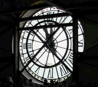
Telescopes
Telescopes use two or more lenses to enlarge distant objects.
There are two kinds of refractor telescopes:
- Galilean telescopes which provide an upright image.
- Keplerian telescopes which invert the image. The objective or main lens forms a real image which is magnified by the eyepiece for the observer.
There are four main types of reflector telescopes:
- Newtonian: light reflected from a parabolic mirror back along its path to a small flat mirror, which sends it at right angles to the main telescope tube and out to the eyepiece.
- Herschel: light is reflected from a parabolic mirror back at an angle through an eyepiece.
- Cassegrain: light reflected from a parabolic mirror is turned back on its path a third time and sent through a hole in the main mirror to an eyepiece.
- Coude: like the Cassegrain, except that instead of passing through a hole in the primary mirror, the light is reflected by a third mirror out at right angles to the main telescope tube, and can be used by massive equipment which must be fixed in place.
In all cases, the point is to increase the size of the primary mirror or lens in order to increase the light-gathering capabilities of the telescope and to increase the focal length of the telescope and thus the magnification power. Bending the light back on itself increases the focal length without increasing the size of the telescope--a distinct advantage for reflectors over refractors.
The magnification from a telescope is determined by the ratio of the focal lengths of the objective lens or mirror, and the eyepiece:
If you want more details on how to choose or make telescopes, check out the Amateur Telescope Makers site.
Compound microscopes
The general, overall magnification of a microscope is given by the product of the magnifications of the objective and eyepiece. On most microscopes, the eyepiece and objectives can be varied. For example, I have two eyepieces, 10X and 20X, for my microscope, and three objectives, 8X, 10X, and 20X. The maximum magnification is thus 20 X * 20X, or 400X.
The magnification of the eyepiece (which acts like a simple lens) will be
For most people, the near point N will be about 25 cm.
The magnification of the of the objective will be
But the image distance must be the length between the objective and eyepiece less the focal length of the eyepiece, l - fe, in order to put the image at the focal point of the eyepiece. So
Now, if fo and fe are very small, then l - fe ≈ fe, and fo ≈ do. Substituting these magnifications back into M (overall magnification) = Mo*Me reduces to
Lens aberrations
Aberrations or variation in focal length occurs in the light passing through a lens for several reasons.
Spherical aberration occurs because light doesn't normally strike a lens in perfect parallel rays, so the rays which strike the edge of the lens at an angle don't focus at the same point as the rays which strike the center of the lens perpendicular to the surface of the lens.
Coma occurs when an image doesn't lie symmetrically on the lens axis. Then a kind of spherical aberration occurs to the light coming from different parts of the object. Distortion occurs when different parts of the object are at different distances from the lens (similar to the problem of keeping everything in the field of view in focus for a camera). Chromatic aberration occurs because different colors of light bend different amounts; since blue "bends better", its focal point will lie closer to the lens than red light's focal point. A colored object thus appears blurred.
Resolution
Resolution is the ability of a lens to separate two close points into separate images. Aberrations which blur the image will make it difficult for a lens to accomplish this; so will the phenomena of diffraction. Since the lens has edges, light will bend around the edge of the lens and into the image area, with the same maxima and minima bands we have already studied for slit diffraction.
The central image of a diffraction pattern from a point source of light falling on a lens has a maximum angular width which is a function of the wavelength of the light and the diameter of the lens:
Compare this to the relationship between the width of a single slit and the first minima where m = 1, of light of wavelength lambda passing through the slit. The factor is 1.22 instead of 1 because we must consider the "average" width of a circular hole (or lens), rather than the uniform width of a slit.
The Rayleigh criterion
The Rayleigh criterion states the resolution limit due to diffraction for a lens of diameter D. Two objects will be resolvable into two images when θ = 1.22 * λ / D--which makes sense, because the first minima of each object's diffraction pattern then lies across the other object's maxima, and the division between them is delineated.
Telescope/Microscope resolution
The resolving power of a set of lenses, whether in a microscope or a telescope, is a result of the Rayleigh criterion. We define the resolving power RP as the distance s between two close objects. For a microscope, when the object distance is close to the focal length of the lens, the distance s = f * θ, where thetatheta; is the angle between the object position and the edges of the magnifying lens. RP is then 1.22 * lambda * f/D
We sometimes consider the angle of acceptance alpha instead, defining it to be half of θ. The resolving power is then expressed as RP = 0.61 λ/sin α.
Human eye resolution
There is a limit to the ability of the human eye to resolve images, which of course varies with the wavelength of the light. This limitation puts a practical limit to the magnification achievable by microscopes at about 500X.
X-rays and X-ray diffraction
While limiting diffraction can be a frustrating exercise for the biologist intent on getting a clear image of cell organelles, it can be a useful phenomenon for the physicist. By bouncing x-rays off of crystals and measuring the diffraction patterns, x-ray crystallographers have mapped the location of specific atoms in molecules, from simple minerals to complex proteins.
Practice with the Concepts
Discussion Points
- Why is the depth of field greater, and the image sharper, when a camera lens is “stopped down” to a larger f-number? Ignore diffraction. horizontal to obtain best reception?
- In attempting to discern distant details, people will some- times squint. Why does this help?
© 2005 - 2026 This course is offered through Scholars Online, a non-profit organization supporting classical Christian education through online courses. Permission to copy course content (lessons and labs) for personal study is granted to students currently or formerly enrolled in the course through Scholars Online. Reproduction for any other purpose, without the express written consent of the author, is prohibited.